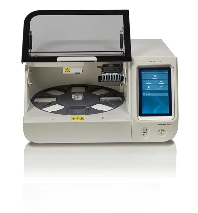
Thromboelastography (TEG) is an emerging technology that provides a novel method for analyzing the entire coagulation process from initial fibrin formation through fibrinolysis. By continuously monitoring clot formation and strength over time, TEG gives physicians deeper insights into a patient’s coagulation status compared to conventional coagulation tests. With its ability to detect hypercoagulable states as well as bleeding risks, TEG is gaining increasing recognition as a valuable tool in transfusion medicine and perioperative care.
What is Thromboelastography?
TEG analyzes whole blood coagulation by measuring the viscoelastic properties of clot formation and breakdown. In a TEG test, whole blood is placed in a cup and rotated back and forth. As the cup oscillates, a pin suspended in the cup detects the formation, strength and breakdown of the blood clot over time. A computer records and analyzes the pin motion generating a thromboelastogram tracing the entire coagulation process from initial fibrin formation through fibrinolysis. Several parameters can be derived from the tracing providing a comprehensive assessment of hemostasis.
Parameters Measured by TEG
R time measures the initiation of clot formation reflecting the time taken for initial fibrin formation. Prolonged R times indicate coagulation factor deficiencies. K time reflects the time taken to achieve a specified clot firmness once clot formation begins. Prolonged K times suggest impaired rate of clot development. Angle α measures the speed of clot strengthening or kinetics of fibrin build-up and cross-linking. A decreased α suggests impairment of clot development. Maximum Amplitude (MA) indicates maximum clot strength achieved and sensitivity to fibrinolysis. A decreased MA suggests decreased clot strength. Lysis 30 and Lysis 60 measure the percentage of clot dissolution or lysis at 30 and 60 minutes post MA. Increased lysis indicates hyperfibrinolysis.
Applications of Thromboelastography
TEG provides a more comprehensive global assessment of hemostasis compared to conventional coagulation tests which measure individual components of the clotting cascade. Its ability to detect coagulation factor deficiencies as well as abnormalities of platelet number and function makes it especially useful in perioperative and trauma settings where massive transfusions are often required. TEG guided transfusion protocols minimizing blood product use have been shown to reduce costs and improve outcomes in cardiac surgery, liver transplantation and trauma. Its ability to detect hypercoagulable states also aids in management of thrombotic conditions like cerebral venous sinus thrombosis. TEG is emerging as a key technology for personalized hemostatic management in high risk patients.
Assessing Coagulopathy of Liver Disease
Liver disease poses major bleeding and clotting risks due to compromised synthesis of coagulation proteins and platelets. Conventional coagulation tests often fail to accurately predict bleeding in liver patients due to their inability to evaluate fibrinogen and platelet function. TEG provides a more reliable assessment of the coagulation status in liver disease, detecting both bleeding risks as well as prothrombotic tendencies. Multiple studies have found TEG parameters to correlate better with bleeding compared to conventional tests in liver patients undergoing invasive procedures or suffering variceal hemorrhage. TEG guided transfusion algorithms in liver transplantation reduce transfusions without increasing bleeding.
Detecting Trauma Induced Coagulopathy
Trauma induced coagulopathy is a major cause of mortality in severely injured patients. Standard coagulation tests lack the sensitivity to detect the early development of trauma coagulopathy characterized by impairment of clot formation and strength. TEG identifies trauma coagulopathy within 30 minutes of admission, much earlier than conventional tests, allowing rapid goal directed resuscitation and correction of hemostatic abnormalities. TEG guided management incorporating focused correction of clotting factor deficiencies, fibrinogen replacement and platelet transfusions according to TEG parameters has been shown to reduce blood product use and improve survival in trauma patients requiring massive transfusions.
Hypercoagulable States and Thrombosis
Inherited and acquired hypercoagulable conditions increase thrombotic risks. Conventional coagulation tests do not reliably distinguish between bleeding risks and hypercoagulable tendencies. TEG parameters like decreased R and K times, increased α angle and MA reflecting accelerated clot formation and strength can aid in identifying hypercoagulable etiologies in patients with thrombosis. A meta-analysis found TEG MA to have a sensitivity of 74% and specificity of 76% in detecting Factor V Leiden, the most common cause of inherited hypercoagulability. Studies also report TEG abnormalities in antiphospholipid syndrome and paroxysmal nocturnal hemoglobinuria indicating a hypercoagulable state. Thus TEG aids in coagulation work-up of thrombophilic conditions.
In conclusion, thromboelastography provides a global analysis of hemostasis from initial fibrin formation through clot lysis enabling a more comprehensive understanding of coagulation compared to standard coagulation tests. With its emerging applications in transfusion guidance, management of coagulopathies and detection of bleeding and hypercoagulable risk states, thromboelastography is growing in clinical importance as a personalized approach to hemostatic management. Larger prospective trials are still needed to further define optimal transfusion algorithms based on TEG parameters. With technological improvements enhancing accessibility, thromboelastography holds much promise as a valuable addition to the coagulation laboratory.


