 by
by Introduction
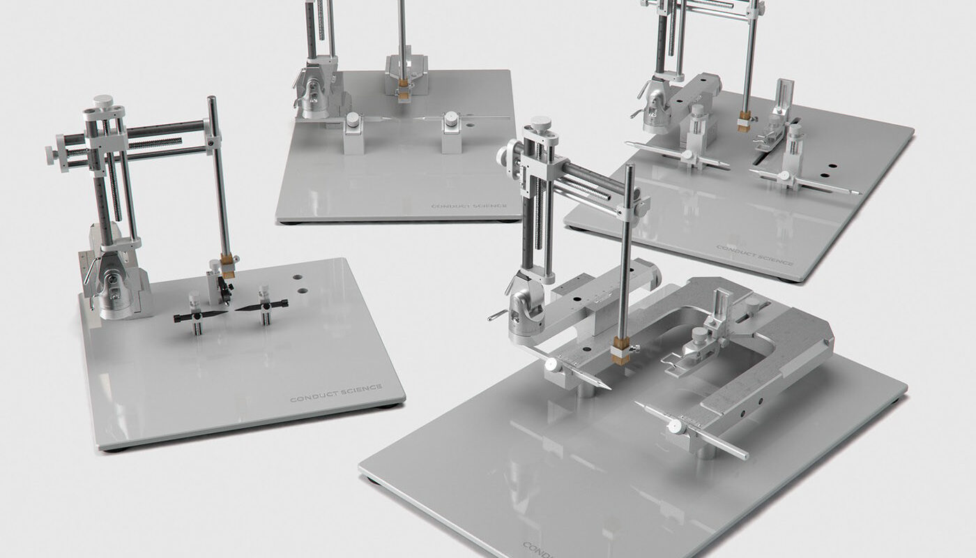
Stereotaxic instruments, also known as stereotactic frames, are precision devices used for targeting specific sites within the brain. They allow for accurate localization and manipulation of brain structures in laboratory animals and have played an important role in advancing our understanding of neuroanatomy and neurophysiology. In this article, we will discuss the history and development of stereotaxic instruments, how they work, applications in research, and their impact on neuroscience.
Historical Development
The origins of stereotaxic instruments can be traced back to the late 19th century when pioneering neurosurgeons began experimental operations on the brains of animals. In the 1890s, German anatomist Korbinian Brodmann created one of the first atlases of the primate brain, dividing it into 52 regions based on cytoarchitecture. This early work laid the foundation for mapping the structure and function of the brain.
In the early 1900s, physicians Ernest Spiegel and Henry T. Wyckoff developed stereotactic frames for precisely locating tumors in human patients. Meanwhile, researchers including Robert Yerkes and James W. Papez used simple stereotaxic devices to study the functions of different brain regions through electrical stimulation in animals. Throughout the 1900s, stereotaxic instruments became more sophisticated with the integration of microelectrodes and other tools, aiding neurological research.
How They Work
All Stereotaxic Instruments work on the principle of coordinate systems and reference planes to localize brain areas. The device secures the animal’s head in a fixed position. Surgical tools can then be guided to target locations based on their relative position to calibrated reference points. Early frames used anatomical landmarks like bregma, a point where the sagittal and coronal sutures intersect. More advanced instruments integrate MRI and CT scans to create digital atlases for three-dimensional targeting. Microelectrodes, injectors and other devices fit onto manipulators that interface with the frame’s coordinate system.
Neurophysiology
Stereotactic instruments enabled systematic mapping of sensory and motor functions in the rodent brain through single-unit recording and electrical microstimulation. Landmark studies in the 1950s mapped somatosensory and motor cortices, helping establish their functional organization. More recent work has elucidated the neural circuitry underlying complex behaviors like learning and memory by recording neuron activity across brain areas.
Neuroanatomy
The precision targeting afforded by stereotactic frames is ideal for neuroanatomical tract-tracing experiments. By injecting neuronal tracers into defined brain nuclei, their efferent and afferent connections can be elucidated. This has advanced our understanding of large-scale brain circuits involved in sensory, motor and higher-order functions. Stereotactic surgery also facilitates lesion studies to determine the functional significance of specific brain regions.
Neuropharmacology
Precise drug delivery via microinjection electrodes and cannulae has helped uncover the neurochemical bases of behavior. For example, local application of receptor agonists/antagonists paired with behavioral testing illuminated the roles of neurotransmitters like dopamine and serotonin. Additionally, stereotactic gene delivery techniques enable manipulation and tracing of genetically-defined neuronal populations. Together, these approaches have revolutionized our knowledge of neurochemistry.
Impact on Our Understanding of the Brain
There is no doubt stereotactic methods have catalyzed progress across neuroscience fields. By enabling targeted experiments down to the level of individual neurons and neurotransmitter systems, they have helped build sophisticated models of information processing in the brain. Key advances include elucidating cortical functional maps, discovering circadian and reward pathways, mapping learning and memory circuits, and deciphering the underpinnings of neurological and psychiatric disorders. Stereotaxis remains an essential toolset for both basic and translational investigation into brain structure and function. Future applications may include multi-electrode arrays, optogenetics and spatial transcriptomics to probe neural computation across scales. Stereotactic approaches will continue advancing our knowledge of the brain’s intricate workings.
In this article, we discussed the origins and evolution of stereotaxic instruments from early anatomical mapping tools to today’s digital platforms. Their ability to precisely localize brain structures has empowered landmark neurophysiological, neuroanatomical and neuropharmacological discoveries. By targeting specific regions and cell types, stereotactic methods have been integral to unraveling how the brain gives rise to cognition, emotion, sensation and movement. Looking ahead, refinements in engineering and multi-disciplinary applications ensure stereotaxis will remain a driving force pushing the boundaries of neuroscience towards a deeper mechanistic understanding of both healthy and diseased brain function.
*Note:
1. Source: Coherent Market Insights, Public sources, Desk research
2. We have leveraged AI tools to mine information and compile it