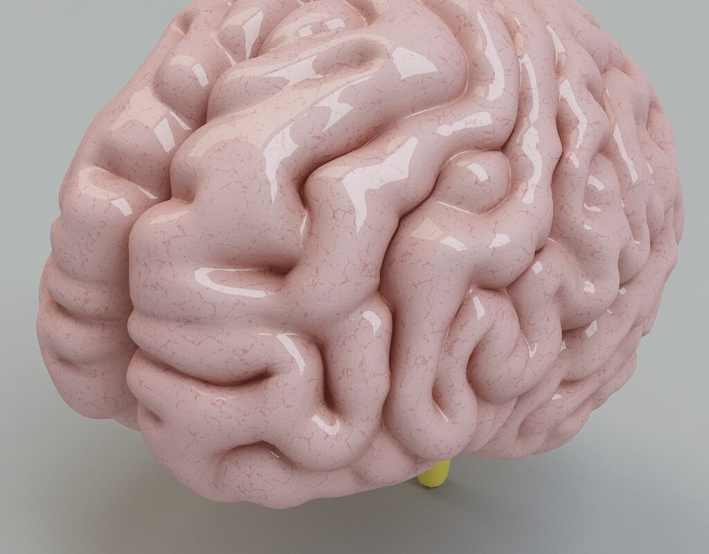
Scientists at the University of Wisconsin–Madison have achieved a significant milestone in the field of neuroscience by successfully developing 3D-printed brain tissue that functions like natural brain tissue. This breakthrough has profound implications for the study and treatment of neurological and neurodevelopmental disorders such as Alzheimer’s and Parkinson’s disease.
The team, led by Professor Su-Chun Zhang of UW–Madison’s Waisman Center, believes that this 3D-printed brain tissue could offer valuable insights into the communication and interaction between brain cells and different parts of the brain. Zhang describes it as a potentially revolutionary tool that could transform our understanding of stem cell biology, neuroscience, and the underlying causes of various neurological and psychiatric conditions.
Previous attempts at printing brain tissue were hampered by limitations in printing methods. By taking a different approach in their study, the researchers at UW–Madison were able to overcome these obstacles. Instead of stacking layers vertically, they decided to arrange the brain cells horizontally using a softer bio-ink gel.
The result is a tissue that possesses enough structure to hold itself together while remaining soft enough to allow the neurons to grow and connect with each other. The neurons are positioned next to each other in a manner similar to pencils laid on a table.
One of the advantages of this new printing technique is that the tissue remains relatively thin, which helps the neurons receive sufficient oxygen and nutrients from the growth media. The cells are able to form connections within each printed layer and across different layers, forming networks that resemble those found in natural human brains. The neurons communicate, send signals, interact through neurotransmitters, and even form networks with support cells that were added to the printed tissue.
In their experiments, the researchers successfully printed sections of the cerebral cortex and the striatum and were amazed by the results. Even when different cells from different parts of the brain were printed, they were still able to communicate in a distinct and significant manner.
Compared to brain organoids, which are miniature organs used to study brains, the 3D-printed tissue offers a higher level of precision and control over the types and arrangement of cells. Organoids tend to grow with less organization and control.
Zhang highlights the unique capabilities of his lab, emphasizing their ability to produce various types of neurons at any time and arrange them in any desired way. This flexibility allows them to closely examine how the human brain network functions under specific conditions.
By printing the tissue according to their design, the researchers can focus on studying how nerve cells communicate with each other in precise ways. This level of control enables them to gain insights into the intricate workings of the human brain.
The groundbreaking research on 3D-printed functional brain tissue could potentially revolutionize the field of neuroscience, leading to new discoveries and advancements in our understanding and treatment of brain-related disorders.
*Note:
- Source: Coherent Market Insights, Public sources, Desk research
- We have leveraged AI tools to mine information and compile it


