Researchers from the Universities of Surrey and Oxford have made a significant discovery regarding the risks associated with cell therapies for heart repair. The study found that a specific type of cell, called myofibroblasts, which are crucial in tissue repair after a heart attack, may also contribute to an increased risk of rhythm disorders associated with cutting-edge cell therapies. This breakthrough could pave the way for safer regenerative treatments for individuals who have suffered a heart attack.
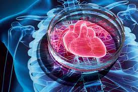
The study focused on the interactions between two types of cells: Human induced pluripotent stem cell-derived cardiomyocytes (hiPSC-CMs), which are created in the lab from stem cells, and myofibroblasts, which are responsible for repairing heart tissue after a heart attack.
Published in Cellular and Molecular Life Sciences, the study revealed that myofibroblasts affect the electrical properties and calcium handling of hiPSC-CMs. Additionally, these cells altered the expression of genes responsible for vital functions of heart cells, which caused electrical instability.
Dr. Patrizia Camelliti, the lead author of the study from the University of Surrey, emphasized the importance of understanding the relationship between myofibroblasts and hiPSC-CMs in order to develop safe regenerative treatments for heart attack patients. The study identified Interleukin-6 (IL-6), a molecule released by myofibroblasts involved in inflammatory responses, as a key player in this interaction. The research team found that blocking IL-6 signaling reduced the negative effects of myofibroblasts on heart cells.
While this study provides valuable insights into the unintended consequences of cell therapies on heart rhythm, further research is necessary to translate these findings into clinical practice.
Heart attacks result in the loss of heart muscle cells and the formation of scar tissue, which involves myofibroblasts. To regenerate healthy heart muscles and repair this damage, scientists have been investigating the use of stem cells, such as hiPSC-CMs. However, these therapies have been shown to increase the risk of heart rhythm disorders, which can be life-threatening.
The research team utilized advanced cell culture systems to recreate the interactions between hiPSC-CMs and adult human cardiac myofibroblasts. By observing the effects on heart cell function, they cultured the two cell types in three different conditions to simulate their interactions: direct contact, non-contact, and medium conditioning.
Dr. Patrizia Camelliti also discussed the potential of targeting interactions between cardiac cells as a novel therapeutic strategy to improve the outcomes of cardiac cell therapies and treat heart rhythm disorders. This perspective was published in Science.
This groundbreaking study provides important insights into the risks associated with cell therapies for heart repair. By uncovering the role of myofibroblasts in contributing to heart rhythm disorders, researchers can now focus on developing safe and effective regenerative treatments for individuals who have experienced a heart attack. With further research and advancements, it is hopeful that these findings will ultimately result in improved patient outcomes and a reduced risk of complications.


